
Published: February 16, 2024
Last updated: October 10, 2026
Written and source checked by: Adel Galal, Founder and Lead Writer at NextFitLife
Medical review status: This article has not been medically reviewed by an ophthalmologist, optometrist, neurologist, cardiologist, hematologist, or other licensed healthcare professional.
You look in the mirror and see a patch of blood in your eye. Or your vision suddenly becomes blurry and you wonder whether a blood clot has blocked the eye.
Those two situations are very different.
Quick answer: Blood clots in eye prevention depends on what type of eye blood vessel problem you mean. A red patch on the white of the eye is usually a subconjunctival hemorrhage, which is bleeding from a small surface vessel and is usually harmless. A retinal vein occlusion blocks blood leaving the retina. A retinal artery occlusion blocks blood reaching the retina and is an eye stroke that needs emergency assessment.
The strongest prevention steps focus on blood pressure, diabetes, cholesterol, smoking, cardiovascular health, glaucoma, and appropriate medical follow up.
This article is for general information and education. It does not replace medical advice, diagnosis, or treatment.
Sudden Vision Loss Is an Emergency
If vision suddenly disappears, becomes severely blurred, or part of the visual field suddenly goes missing in one eye, do not wait for the symptom to settle.
Central retinal artery occlusion is considered an acute ischemic stroke involving the retina. The American Heart Association and ophthalmology guidance recommend immediate emergency evaluation.
Get urgent medical help for:
- Sudden painless loss of vision in one eye
- Sudden major blurring in one eye
- A sudden missing area of vision
- Temporary sudden loss of vision that returns
- Sudden vision change with weakness, facial drooping, or speech difficulty
- Sudden vision loss in a person over 50 with a new severe headache, scalp tenderness, or jaw pain
Do not drive yourself if vision has suddenly changed.
A bright red patch on the white of the eye with normal vision and no significant pain is a different pattern and is commonly caused by a surface hemorrhage.
Table of Contents
- What Blood Clot in the Eye Means
- Surface Bleeding vs Retinal Blockage
- 12 Smart Prevention Steps
- Blood Pressure and Eye Vessels
- Diabetes and Cholesterol
- Smoking and Cardiovascular Risk
- Glaucoma and Eye Pressure
- Aspirin and Blood Thinners
- Supplements and Natural Blood Thinners
- What Happens After an Eye Clot
- When Eye Redness Is Usually Harmless
- Frequently Asked Questions
What Does “Blood Clot in the Eye” Actually Mean?
The phrase is not a precise diagnosis.
People commonly use it for at least three different problems.
| Condition | What Happens | Typical Clue |
|---|---|---|
| Subconjunctival hemorrhage | A tiny surface vessel breaks and leaks blood under the clear covering of the eye | Bright red patch with normal vision and usually no pain |
| Retinal vein occlusion | A retinal vein becomes blocked, often by a clot | Painless blurred or reduced vision in one eye |
| Retinal artery occlusion | Blood flow through a retinal artery stops, often because of an embolus or thrombus | Sudden painless vision loss and a medical emergency |
That distinction changes both treatment and prevention.
For a detailed symptom guide, read Blood Clot in Eye: Critical Signs and Safe Care.
Why Can Retinal Blood Vessels Become Blocked?
The retina needs a steady blood supply.
In retinal vein occlusion, blood cannot drain normally from part or all of the retina.
The National Eye Institute lists important risk factors for central retinal vein occlusion such as:
- High blood pressure
- Diabetes
- Glaucoma
- Atherosclerosis
- Older age
Smoking, cardiovascular disease, high cholesterol, obesity, and selected blood disorders can also contribute to retinal vascular risk.
For retinal artery occlusion, risk factors overlap strongly with stroke risk.
Possible sources of an embolus include the carotid arteries and the heart.
That is why an eye stroke can lead to an urgent cardiovascular and neurological evaluation rather than only an eye examination.
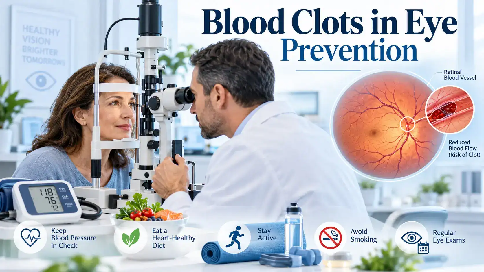
12 Smart Steps for Blood Clots in Eye Prevention
1. Control High Blood Pressure
This is one of the most important modifiable risk factors.
Long term high blood pressure damages blood vessel walls throughout the body, including the small vessels in the retina.
It is strongly linked with retinal vein occlusion and retinal artery disease.
Know your blood pressure rather than assuming it is normal because you feel well.
If it is high, follow the treatment plan recommended by your healthcare professional.
That may include:
- Prescribed medicine
- Less excess sodium
- Regular activity
- Weight management
- Limiting heavy alcohol use
- Regular blood pressure checks
For more help, read High Blood Pressure and What Causes High Blood Pressure?.
2. Manage Diabetes
High blood glucose can damage small blood vessels over time.
Diabetes is associated with retinal vein occlusion and several other eye problems.
The useful goal is not simply “avoid sugar.”
Work with your healthcare team on:
- Blood glucose targets
- A1C when appropriate
- Blood pressure
- Cholesterol
- Medicines
- Eye examinations
Visit Eye Health and Diabetes and the Diabetes and Blood Sugar Management Hub.
3. Treat High Cholesterol and Atherosclerosis Risk
High cholesterol does not literally make blood “sticky.”
The more accurate issue is that unhealthy cholesterol patterns contribute to atherosclerosis, where plaque builds inside arteries.
That matters because retinal artery occlusion can result from material traveling from diseased arteries into the retinal circulation.
Diet, activity, smoking cessation, weight management, and cholesterol lowering medicine when prescribed all have roles.
See High Cholesterol and the Heart and Cardiovascular Health Hub.
4. Stop Smoking
Smoking damages blood vessels and raises cardiovascular risk.
Retinal artery and retinal vein disease share many of the same risk factors as heart disease and stroke.
If you smoke, quitting is one of the strongest steps you can take for both your eyes and your wider vascular health.
This matters more than buying an eye supplement.
Read Smoking Eye Damage for the wider effects of tobacco on vision.
5. Stay Physically Active
Exercise does not flush clots out of the retina.
Its value is broader.
Regular activity supports:
- Blood pressure
- Blood sugar
- Cholesterol
- Body weight
- Heart health
Those are the same systems connected with retinal vascular disease.
Choose activity that fits your fitness level and medical conditions.
6. Work Toward a Healthy Body Weight
Obesity is linked with vascular disease and is listed among risk factors for retinal artery occlusion.
Weight reduction can also improve blood pressure, glucose control, and cardiovascular risk when excess body weight is present.
You do not need rapid weight loss.
A sustainable change in eating and activity is more useful.
7. Check for Glaucoma and High Eye Pressure
Glaucoma is an important risk factor for retinal vein occlusion.
You cannot reliably feel whether your eye pressure is high.
Regular eye care becomes more important if you already have glaucoma or are at increased risk.
If glaucoma is diagnosed, use prescribed eye drops and attend follow up visits as directed.
Do not stop glaucoma treatment because the eye feels normal.
8. Keep Eye Examinations Appropriate to Your Risk
Routine eye examinations do not guarantee that a retinal clot will never happen.
They can still identify conditions that matter, including:
- Glaucoma
- Diabetic retinal changes
- High eye pressure
- Existing retinal disease
People with diabetes usually need regular dilated retinal assessment according to their diabetes care plan.
Visit the Eye Health Complete Guide for the wider vision care plan.
9. Treat Heart Rhythm and Carotid Problems Properly
This is especially important for retinal artery occlusion.
A clot or embolus can travel to the retinal artery from elsewhere in the circulation.
Important conditions can include:
- Atrial fibrillation
- Carotid artery disease
- Heart valve disease
- Other cardiovascular disease
If one of these conditions has been diagnosed, follow the prescribed treatment plan.
Do not alter heart or anticoagulant medicine because of an eye article.
10. Take Prescribed Antiplatelet or Anticoagulant Medicine Correctly
Blood thinning medicines can be lifesaving when a person has the right medical reason to take them.
They are not a universal eye clot prevention treatment.
After a retinal artery occlusion, doctors may recommend antiplatelet or anticoagulant therapy depending on the underlying stroke or cardiovascular cause.
That decision can depend on conditions such as atrial fibrillation, carotid disease, prior stroke, bleeding risk, and other medical history.
Do not start aspirin, warfarin, heparin, or another anticoagulant just because you are worried about an eye clot.
Do not stop one that has been prescribed without speaking with the clinician who manages it.
11. Avoid DIY “Natural Blood Thinning”
This is a major correction to the older version of this article.
Fish oil, ginkgo, garlic, ginger, turmeric, cayenne, and other supplements should not be used as homemade anticoagulant therapy for retinal vessel occlusion.
Some supplements can affect bleeding or interact with prescription medicine.
Even if a supplement changes platelet activity in a laboratory study, that does not prove it prevents retinal artery or vein occlusion in real people.
Prevention should target proven cardiovascular and eye risk factors.
12. Review Repeated Surface Eye Bleeding Instead of Calling It a Clot
A subconjunctival hemorrhage often appears as a dramatic bright red patch.
It usually does not hurt or change vision.
Common triggers can include:
- Coughing
- Sneezing
- Vomiting
- Straining
- Eye rubbing
- Minor injury
High blood pressure, diabetes, anticoagulant medicines, and bleeding disorders can increase risk.
If the bleeding keeps returning, discuss it with a healthcare professional. Blood pressure, medicine use, and clotting problems may need review.
A recurrent red patch is not a reason to stop prescribed blood thinners on your own.
Does Drinking More Water Prevent Eye Blood Clots?
Normal hydration is good for general health.
But “drink more water so your blood cannot become thick and sticky” is too simple and is not a proven retinal clot prevention strategy.
You do not need to force large amounts of water.
Focus first on the better established risks:
- Blood pressure
- Diabetes
- Cholesterol
- Smoking
- Glaucoma
- Cardiovascular disease
People with heart failure, kidney disease, or a prescribed fluid restriction should follow their own medical fluid plan.
Do Screen Breaks Prevent Blood Clots in the Eye?
No evidence shows that taking breaks from reading, phones, or computer work prevents retinal artery or vein occlusion.
Screen breaks can help with digital eye strain, dryness, and focusing discomfort.
That is a different problem.
Read Digital Eye Strain Relief if screen discomfort is your concern.
Can Eye Surgery Cause Blood Clots in the Eye?
Do not assume that every person who has eye surgery is suddenly at high risk for a retinal blood clot.
Different operations have different complications and recovery instructions.
The useful advice after eye surgery is to follow the surgeon's specific plan and report unexpected vision loss, severe pain, flashes, a sudden shower of floaters, or other major visual changes promptly.
The older version of this article treated eye surgery as a general retinal clot risk and suggested measures such as head elevation and near work breaks as clot prevention. That is too broad.
What Is a Retinal Vein Occlusion?
A retinal vein occlusion happens when a vein carrying blood away from the retina becomes blocked.
There are two main types:
- Central retinal vein occlusion, or CRVO, involving the main retinal vein
- Branch retinal vein occlusion, or BRVO, involving a smaller branch
Symptoms commonly include blurry or reduced vision in one eye.
Some mild cases can initially have few symptoms.
The National Eye Institute identifies high blood pressure, diabetes, glaucoma, arteriosclerosis, and older age as important CRVO risks.
Can a Retinal Vein Occlusion Be Dissolved With a Blood Thinner?
Not in the simple way many people imagine.
Standard eye treatment focuses on complications caused by the occlusion.
One major complication is macular edema, where fluid collects in the central retina.
Treatment can include:
- Anti VEGF injections
- Selected steroid treatment
- Laser treatment in certain complications
The eye doctor also looks for conditions such as glaucoma.
At the same time, general vascular risk factors need to be identified and managed.
What Is a Retinal Artery Occlusion?
A retinal artery occlusion blocks the blood supply going into the retina.
The central form is called central retinal artery occlusion, or CRAO.
The American Heart Association defines CRAO as a form of acute ischemic stroke.
It often causes sudden, painless loss of vision in one eye.
The obstruction is commonly caused by material that travels through the bloodstream and lodges in a retinal artery.
Potential sources include the carotid arteries and heart.
This is why the patient may need:
- Stroke evaluation
- Heart rhythm assessment
- Carotid assessment
- Blood pressure testing
- Cholesterol testing
- Diabetes assessment
Read Stroke Symptoms for the wider emergency warning signs.
Why Is Giant Cell Arteritis Important?
In people over age 50, retinal artery blockage can sometimes be related to giant cell arteritis, an inflammatory disease of medium and large arteries.
Warning symptoms can include:
- A new headache
- Scalp tenderness
- Jaw pain while chewing
- Sudden vision changes
- Feeling generally unwell
This requires urgent medical assessment because the other eye can also be threatened.
Is a Red Patch on the White of the Eye a Blood Clot?
Usually, no.
A bright red patch on the white of the eye is most commonly a subconjunctival hemorrhage.
A small blood vessel has broken under the clear conjunctiva.
Mayo Clinic notes that it usually causes:
- No vision change
- No discharge
- No significant pain
The blood is gradually absorbed, often over days to a few weeks.
Artificial tears may help mild irritation, but eye drops do not need to “dissolve the blood.”
When Should a Red Eye Be Checked?
Get medical advice if:
- Vision changes
- The eye is painful
- The redness followed an injury
- Bleeding happens repeatedly
- You also bruise or bleed unusually elsewhere
Sudden visual loss is a very different situation from an isolated painless surface red patch and needs emergency assessment.
Should You Stop Aspirin if the White of Your Eye Turns Red?
Not on your own.
Aspirin and anticoagulants can make a surface hemorrhage more likely or more noticeable, but they may have been prescribed to prevent serious stroke, heart, or clotting problems.
Stopping them without medical advice can create a larger risk than the eye redness itself.
If hemorrhages repeatedly occur, ask the prescribing clinician whether your medicine and dose still fit your health needs.
Can Eye Vitamins Prevent Retinal Blood Clots?
There is no vitamin proven to prevent retinal vein or retinal artery occlusion simply by taking it as a supplement.
A nutritious diet supports cardiovascular and general eye health, but supplements do not replace:
- Blood pressure control
- Diabetes care
- Cholesterol treatment
- Smoking cessation
- Glaucoma care
- Cardiovascular treatment
For evidence based nutrition guidance, see Vitamins for Eyes.
What Happens After a Retinal Vessel Occlusion?
Prevention becomes even more important after one event.
The medical team may investigate risk factors that affect both eyes and the rest of the circulation.
This can include:
- Blood pressure
- Blood sugar
- Cholesterol
- Smoking
- Glaucoma
- Heart disease
- Atrial fibrillation
- Carotid artery disease
- Selected clotting disorders
After retinal artery occlusion, secondary stroke prevention may require collaboration between ophthalmology, neurology, primary care, and cardiovascular specialists.
After retinal vein occlusion, eye follow up matters because complications such as macular edema, abnormal new blood vessels, or glaucoma can affect vision.
Do Young Adults Need Blood Clotting Tests?
Not every person with retinal vein occlusion needs an enormous clotting panel.
Younger patients, people with an unusual presentation, or those without the common vascular risk factors may need investigation for less common causes.
That decision belongs with the ophthalmologist and medical team.
Testing everyone for every rare clotting disorder can produce confusing results without improving care.
A Simple Eye Vessel Risk Checklist
| Risk Area | Useful Action |
|---|---|
| Blood pressure | Know your numbers and treat persistent hypertension |
| Diabetes | Manage glucose and attend recommended retinal screening |
| Cholesterol | Treat cardiovascular risk based on your medical plan |
| Smoking | Stop tobacco exposure |
| Glaucoma | Use prescribed treatment and attend follow up |
| Heart rhythm | Treat atrial fibrillation according to medical advice |
| Medicines | Do not start or stop anticoagulants on your own |
| Sudden vision loss | Seek emergency assessment immediately |
Common Eye Clot Prevention Mistakes
Calling Every Red Patch a Clot
A surface hemorrhage and a retinal vessel occlusion are different conditions.
Taking Aspirin Without Medical Advice
Aspirin has real bleeding risks and should be used for the right medical reason.
Using Supplements to Thin the Blood
Garlic, ginkgo, turmeric, ginger, fish oil, and similar products are not proven retinal clot prevention treatments.
Thinking Water Alone Prevents Clots
Normal hydration matters, but vascular risk control matters much more.
Taking Screen Breaks to Improve Retinal Circulation
Screen breaks help eye strain, not retinal clot prevention.
Ignoring Glaucoma
Glaucoma is an important risk factor for retinal vein occlusion.
Waiting for Sudden Vision Loss to Improve Overnight
Retinal artery occlusion is an emergency.
Conclusion
Blood clots in eye prevention starts with understanding what you are trying to prevent.
A painless red patch on the white of the eye is usually surface bleeding, not a blocked retinal vessel. Retinal vein occlusion is a deeper vascular problem, while retinal artery occlusion is an eye stroke that requires emergency assessment.
The most useful prevention plan is not a supplement list. Control blood pressure, diabetes, cholesterol, smoking, weight, glaucoma, and cardiovascular disease. Take prescribed medicines correctly and keep appropriate eye follow up.
Your next step is simple: review your blood pressure, glucose, cholesterol, smoking status, eye pressure, and cardiovascular history. If your vision ever changes suddenly in one eye, seek emergency care rather than trying a home remedy.
References and Sources
- National Eye Institute. Central Retinal Vein Occlusion.Federal eye health guidance explaining CRVO symptoms, major risk factors, diagnosis, macular edema, anti VEGF injections, steroids, laser treatment, and follow up.https://www.nei.nih.gov/eye-health-information/eye-conditions-and-diseases/central-retinal-vein-occlusion-crvo
- American Heart Association. Management of Central Retinal Artery Occlusion.Scientific guidance identifying central retinal artery occlusion as an acute ischemic stroke and emphasizing emergency triage, cardiovascular evaluation, and secondary prevention.https://professional.heart.org/en/science-news/management-of-central-retinal-artery-occlusion/top-things-to-know
- American Academy of Ophthalmology EyeWiki. Retinal Artery Occlusion.Ophthalmology guidance on retinal artery occlusion, stroke center referral, causes, risk factors, embolic sources, and emergency assessment.https://eyewiki.aao.org/Central_Retinal_Artery_Occlusion
- MedlinePlus. Retinal Artery Occlusion.NIH and National Library of Medicine guidance covering carotid disease, atrial fibrillation, hypertension, diabetes, cholesterol, diagnosis, prevention, and urgent sudden vision loss.https://medlineplus.gov/ency/article/001028.htm
- MedlinePlus. Retinal Vein Occlusion.Information on retinal vein blockage, vascular risk reduction, diabetes, smoking, weight, complications, and sudden vision change.https://medlineplus.gov/ency/article/007330.htm
- Mayo Clinic. Subconjunctival Hemorrhage.Updated guidance explaining why a red surface patch is usually a broken vessel rather than a retinal clot, plus risk factors, warning signs, and when recurrent bleeding should be assessed.https://www.mayoclinic.org/diseases-conditions/subconjunctival-hemorrhage/symptoms-causes/syc-20353826
Internal Links + Hub
- Eye Health Complete Guide
- Eye Health and Vision Hub
- Blood Clot in Eye: Critical Signs and Safe Care
- Eye Health and Diabetes
- Smoking Eye Damage
- High Blood Pressure
- High Cholesterol
- Heart and Cardiovascular Health Hub
- Stroke Symptoms
- Vitamins for Eyes
About the Author
Adel Galal is the founder and lead writer of NextFitLife. He has maintained a personal interest in health, fitness, vision care, healthy aging, and practical wellness education for more than 30 years, with more than 15 years of health and wellness writing experience.
His work focuses on comparing credible sources, separating established medical guidance from unsafe wellness claims, identifying warning signs, and explaining complex health topics in plain language.
Adel is not a doctor, ophthalmologist, optometrist, neurologist, cardiologist, hematologist, pharmacist, or other licensed healthcare professional. This content does not replace eye examination, emergency stroke assessment, diagnosis, prescription medicine, retinal treatment, or professional medical care.
I am not a dermatologist or a doctor, and this content does not replace professional medical advice. What I share comes from real life experience, extensive research, and consultation with healthcare providers. Always consult qualified medical professionals for diagnosis and treatment of any health condition.
Learn more about Adel, NextFitLife, and its editorial approach on the About NextFitLife page.
Frequently Asked Questions About Blood Clots in Eye Prevention
How can I reduce my risk of blood clots in the eye?
Focus on proven vascular risk factors. Control high blood pressure, diabetes, cholesterol, smoking, excess weight, glaucoma, and cardiovascular disease. Take prescribed medicines correctly and attend recommended eye and medical follow up.
Is a red patch on the white of my eye a blood clot?
Usually not. A painless bright red patch with normal vision is commonly a subconjunctival hemorrhage, where a small surface vessel has broken. It is different from a retinal artery or retinal vein occlusion.
What are the symptoms of retinal vein occlusion?
Retinal vein occlusion often causes painless blurred or reduced vision in one eye. Some mild cases have few symptoms at first. Sudden or unexplained vision change should be assessed by an eye professional promptly.
What are the symptoms of retinal artery occlusion?
Retinal artery occlusion commonly causes sudden, painless loss of vision or a sudden missing area of vision in one eye. Central retinal artery occlusion is an acute ischemic stroke and requires immediate emergency assessment.
Can high blood pressure cause an eye blood vessel blockage?
High blood pressure is one of the major modifiable risk factors for retinal vascular occlusion. Long term hypertension damages blood vessels and is strongly associated with retinal vein disease and retinal artery disease.
Can diabetes increase the risk?
Yes. Diabetes damages small blood vessels and is associated with retinal vein occlusion and other retinal disease. Blood glucose, blood pressure, cholesterol, medicines, and regular retinal care all matter.
Does glaucoma increase the risk of retinal vein occlusion?
Yes. Glaucoma and increased eye pressure are recognized risk factors for retinal vein occlusion. If glaucoma has been diagnosed, use treatment as prescribed and attend follow up visits.
Should I take aspirin to prevent a blood clot in my eye?
Do not start aspirin solely for eye clot prevention without medical advice. Antiplatelet or anticoagulant treatment may be appropriate for specific cardiovascular or stroke related conditions, but it also carries bleeding risk.
Can fish oil, turmeric, garlic, or ginkgo prevent an eye clot?
These supplements are not proven treatments for preventing retinal artery or retinal vein occlusion. Some can affect bleeding or interact with medicines, so they should not be used as homemade blood thinners.
Does drinking lots of water prevent retinal blood clots?
Normal hydration is healthy, but forcing large amounts of water is not a proven retinal clot prevention treatment. Managing blood pressure, diabetes, cholesterol, smoking, glaucoma, and cardiovascular disease has much stronger clinical support.
Are screen breaks useful for preventing eye clots?
No. Screen breaks may reduce dry eye and digital eye strain, but they have not been shown to prevent retinal artery or vein occlusion.
When is an eye blood vessel problem an emergency?
Sudden painless loss of vision, severe sudden blurring, temporary sudden vision loss, or a sudden missing part of the visual field requires urgent assessment. Central retinal artery occlusion is an eye stroke and should be treated as a medical emergency.

Health & wellness writer with 30+ years of experience in nutrition, fitness, and healthy aging. Founder of NextFitLife.com — evidence-based health guidance.