
Published: March 15, 2024
Last updated: October 10, 2026
Written and source checked by: Adel Galal, Founder and Lead Writer at NextFitLife
Medical review status: This article has not been medically reviewed by an ophthalmologist, optometrist, neurologist, retinal specialist, pharmacist, or other licensed healthcare professional.
You may still read clearly, recognize faces, and see what is straight ahead, yet something feels different at the edges of your vision.
You might bump into furniture, miss a person approaching from the side, or notice a dark area that was not there before.
Quick answer: Peripheral vision loss means part of your side visual field is reduced or missing. Gradual loss may occur with glaucoma or inherited retinal disease. Sudden loss can be caused by retinal detachment, stroke, retinal blood flow problems, eye trauma, or another urgent condition. Sudden side vision loss needs prompt medical assessment.
The speed of onset matters. So do flashes, floaters, eye pain, weakness, speech problems, headache, and whether one eye or both visual fields are affected.
This article is for general information and education. It does not replace medical advice, diagnosis, or treatment.
Emergency Warning Signs
Seek urgent eye or emergency care now if you suddenly develop:
- A dark curtain or shadow across your vision
- A sudden shower of new floaters
- Flashes of light with new vision loss
- Sudden loss of side vision in one eye
- Sudden loss of the right or left side of the visual scene
- Sudden major blurring or loss of vision
- Vision loss after significant eye or head trauma
- Severe eye pain with blurred vision
- Severe eye pain with nausea or vomiting
Call emergency services immediately if sudden vision change happens with:
- Facial drooping
- Arm or leg weakness
- Numbness on one side
- Trouble speaking
- Confusion
- Dizziness or loss of balance
- A sudden severe headache
Even if sudden symptoms disappear after a few minutes, they still require urgent assessment because a temporary interruption of blood flow can be a warning sign of a serious vascular problem.
Table of Contents
- What Peripheral Vision Is
- 13 Critical Warning Signs
- Sudden vs Gradual Loss
- Glaucoma
- Retinal Detachment
- Retinitis Pigmentosa
- Stroke and Brain Causes
- One Eye vs Both Eyes
- Visual Field Testing
- Treatment Options
- Can Vision Return?
- Vision Rehabilitation
- Frequently Asked Questions
What Is Peripheral Vision?
Peripheral vision is the part of your visual field outside the point you are looking at directly.
Your central vision helps you read, recognize faces, and see fine detail. Your peripheral vision helps you notice movement, obstacles, people, cars, steps, and objects around you.
Healthy side vision helps with:
- Walking safely through rooms
- Noticing people approaching from the side
- Crossing roads
- Changing lanes
- Avoiding furniture and obstacles
- Moving through crowds
- Playing sports
- Navigating in dim light
A person can keep sharp central vision while losing a meaningful amount of side vision.
This is one reason gradual peripheral vision loss can remain hidden for a long time.
For broader eye guidance, visit the Eye Health and Vision Hub.
13 Critical Signs of Peripheral Vision Loss
1. Bumping Into Doorframes or Furniture
One of the earliest everyday clues is repeatedly clipping the edge of a doorway, table, chair, or other object.
This can happen because the object falls into an area of missing side vision.
One accident means little. A repeated new pattern deserves attention.
2. Missing People Approaching From the Side
You may feel startled when someone appears beside you because you did not see them approaching.
That can happen when part of the outer visual field is reduced.
3. Needing to Turn Your Head More Often
People with side vision loss sometimes compensate without realizing it.
You may begin turning your head more when:
- Walking
- Looking for objects
- Crossing a street
- Shopping
- Following movement
This adaptation can hide the problem for a while.
4. Trouble Finding Things on Crowded Shelves
A busy shelf or table requires you to scan a wide area.
If part of your side vision is missing, objects may seem to disappear unless you look directly at them.
5. Difficulty Driving or Changing Lanes
Peripheral vision helps you detect vehicles, cyclists, pedestrians, and nearby movement.
You may begin to:
- Miss cars beside you
- Feel unsafe changing lanes
- Notice vehicles later than before
- Have trouble seeing at intersections
Do not continue driving after sudden visual field loss.
6. Trouble Walking in Crowded Places
Shopping centers, train stations, airports, and busy streets can become harder to navigate because people and obstacles appear from many directions.
Reduced peripheral awareness may make these spaces feel confusing or unsafe.
7. More Trips or Falls
Missing the edge of a step, curb, rug, or obstacle can increase fall risk.
Vision is not the only cause of falls, but new side vision problems should be considered when collisions or trips begin unexpectedly.
8. Trouble Seeing in Dim Light
Some retinal disorders affect night vision and peripheral vision together.
Retinitis pigmentosa is an important example.
Difficulty adapting to darkness, followed by narrowing side vision, can be an early pattern.
Read Night Vision Problems for other causes of poor vision in darkness.
9. A Narrowing or Tunnel Like View
Some people describe advanced side vision loss as tunnel vision.
This description is useful, but it can also be misleading.
Peripheral loss does not always produce a perfect circular tunnel. Missing areas may be patchy, curved, limited to one side, or affect only part of the upper or lower field.
10. A Curtain or Shadow
A new dark curtain, veil, or shadow is especially important when it begins suddenly.
This is a classic warning pattern for retinal detachment.
If it appears with flashes or many new floaters, seek urgent assessment.
Read Eye Floaters for more information about when floaters become concerning.
11. Missing Half of the Visual Scene
A stroke or brain injury can cause loss of the same side of vision from both eyes.
This pattern is called homonymous hemianopia.
A person may:
- Miss people on one side
- Leave food on one side of a plate
- Miss words at the start or end of a line
- Bump into objects on one side
If this begins suddenly, treat it as an emergency.
12. Flashes With New Floaters
A few long standing floaters are common.
A sudden increase is different.
The combination of flashes, many new floaters, and a missing area of vision can signal a retinal tear or detachment.
13. No Symptoms at All
This may be the most important warning sign of all.
Glaucoma often has no early symptoms.
Side vision can slowly worsen while the person still feels that vision is normal.
This is why screening based only on symptoms is not enough.
Read Glaucoma Prevention and Early Detection.
Sudden vs Gradual Peripheral Vision Loss
| Pattern | Possible Causes | What to Do |
|---|---|---|
| Sudden side vision loss | Retinal detachment, stroke, retinal vascular event, trauma, acute eye disease | Seek urgent medical assessment |
| Slow painless loss | Glaucoma, inherited retinal disease, chronic optic nerve disease | Arrange a comprehensive eye examination |
| Night vision plus narrowing field | Retinitis pigmentosa and other retinal disorders | Retinal specialist assessment |
| Missing same side in both eyes | Brain visual pathway problem, including stroke | Urgent assessment if new or sudden |
What Does Peripheral Vision Loss Look Like?
There is no single appearance.
People may describe:
- Dark areas at the side
- Blurry side vision
- Blind spots
- A narrowing field
- A curtain or shadow
- Missing upper or lower vision
- Missing right or left vision
- Difficulty noticing movement
The popular black tunnel image is only one simplified illustration.
Real visual field defects vary with the cause.
Glaucoma and Peripheral Vision Loss
Glaucoma is one of the most important causes of gradual peripheral vision loss.
It damages the optic nerve, which carries visual information from the eye to the brain.
The National Eye Institute states that open angle glaucoma often begins without symptoms.
As it progresses, side vision may slowly decline.
Important facts include:
- Glaucoma can occur in one or both eyes
- High eye pressure is an important risk factor
- Some people develop glaucoma even with pressure readings in a statistically normal range
- A pressure check alone cannot rule out glaucoma
- A dilated eye exam and visual field test help detect the disease
Can Glaucoma Vision Loss Be Reversed?
Usually not.
Current glaucoma treatment cannot restore optic nerve function that has already been permanently lost.
The goal is to protect remaining vision and slow or stop further damage.
How Is Glaucoma Treated?
Treatment may include:
- Prescription eye drops
- Laser treatment
- Glaucoma surgery
The exact plan depends on the type of glaucoma, eye pressure, optic nerve damage, visual field loss, and other factors.
Retinal Detachment and Sudden Peripheral Vision Loss
Retinal detachment happens when the retina separates from its normal position.
It is an eye emergency.
Warning signs include:
- Many new floaters
- Flashes of light
- A curtain or shadow
- Sudden side vision loss
- Sudden blurring
Early treatment can help protect vision.
How Is Retinal Detachment Treated?
Depending on the problem, treatment can include:
- Laser treatment
- Freezing treatment
- Pneumatic retinopexy
- Scleral buckle surgery
- Vitrectomy
The retinal specialist chooses the approach based on the location, size, and type of detachment.
Retinitis Pigmentosa
Retinitis pigmentosa is a group of inherited retinal diseases.
The National Eye Institute lists loss of night vision and peripheral vision among its early symptoms.
The field can become narrower over time.
Diagnosis may involve:
- Dilated retinal examination
- Visual field testing
- Electroretinography
- Genetic testing
There is no single cure for every form of retinitis pigmentosa.
Management can include low vision aids, genetic counselling, treatment of associated eye problems, and vision rehabilitation.
Can Stroke Cause Peripheral Vision Loss?
Yes.
The eyes may be healthy, but damage to the brain's visual pathways can remove part of the visual field.
Stroke can cause homonymous hemianopia, which means the same right or left half of the visual world is missing from both eyes.
Sudden visual field loss is a stroke warning even if there is no obvious arm weakness.
Other stroke signs can include:
- Face weakness
- Arm or leg weakness
- Numbness
- Speech difficulty
- Confusion
- Balance problems
- Severe sudden headache
Call emergency services if stroke is possible.
Can a Retinal Blood Vessel Problem Affect Side Vision?
Yes.
Blood flow problems in the retina can cause sudden visual loss or missing areas of vision.
Retinal artery blockage is particularly urgent because it can represent an eye and vascular emergency.
Retinal vein blockage can also cause sudden or developing visual change.
Do not try to distinguish these problems at home from the shape of the missing vision.
Can Optic Nerve Problems Cause Peripheral Vision Loss?
Yes.
The optic nerve carries information from the retina to the brain.
Damage can produce different visual field patterns depending on where and how the nerve is affected.
Possible causes include:
- Glaucoma
- Inflammation
- Reduced blood supply
- Compression
- Trauma
- Some nutritional or toxic causes
Optic nerve disease often needs specialist evaluation because treatment depends heavily on the cause.
Can Migraine Cause Temporary Peripheral Vision Changes?
Migraine aura can create temporary visual symptoms.
These can include:
- Flashing lights
- Zigzag patterns
- Shimmering areas
- Temporary blind spots
Do not assume a new sudden visual field defect is migraine, especially if you have never experienced that exact pattern before.
Stroke and retinal disease can mimic visual migraine symptoms.
Does Macular Degeneration Cause Tunnel Vision?
Usually not.
Age related macular degeneration mainly damages the macula, which is responsible for detailed central vision.
It is therefore much more associated with central vision loss than classic tunnel vision.
Read Macular Degeneration Prevention for more information.
Can Cataracts Cause Peripheral Vision Loss?
Cataracts usually cause general blur, glare, faded color, poor contrast, or trouble seeing at night.
They are not a typical cause of a sharply missing peripheral visual field.
That difference matters because a true field defect needs a more targeted eye evaluation.
One Eye vs Both Eyes: Why the Pattern Matters
| Pattern | What Doctors May Consider |
|---|---|
| Sudden loss in one eye | Retinal detachment, retinal vascular problem, optic nerve disease, trauma |
| Gradual loss in one or both eyes | Glaucoma, retinal disease, optic nerve disease |
| Same right or left half missing in both eyes | Brain visual pathway disorder, including stroke |
| Progressive night and side vision loss | Inherited retinal disease such as retinitis pigmentosa |
These patterns guide testing. They do not replace an examination.
How Is Peripheral Vision Tested?
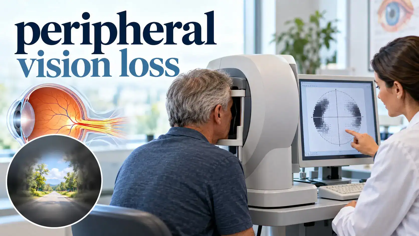
A visual field test maps what you can see while looking straight ahead.
During automated testing, small lights appear in different locations.
You respond when you see them.
The machine then creates a map showing which areas were seen and which were missed.
This is useful for:
- Glaucoma diagnosis
- Monitoring glaucoma progression
- Optic nerve disease
- Retinal disease
- Neurological field loss
What Other Tests May Be Needed?
Dilated Eye Examination
A dilated eye exam allows the eye care professional to inspect the retina and optic nerve more fully.
It is especially important when a retinal tear, retinal detachment, glaucoma, or retinal disease is suspected.
Eye Pressure Testing
Tonometry measures eye pressure.
It is useful for glaucoma evaluation but cannot diagnose or exclude glaucoma on its own.
OCT
OCT creates detailed cross sectional images of structures such as the retina and optic nerve.
It can help identify structural damage that may accompany visual field loss.
Retinal Imaging
Photographs and other retinal scans can document disease and allow change to be tracked over time.
Electroretinography
Electroretinography measures electrical responses from retinal cells.
It can be useful when inherited retinal disease is suspected.
Genetic Testing
Genetic testing can help identify some inherited retinal conditions and guide genetic counselling.
Brain Imaging
CT or MRI may be needed when stroke, compression, trauma, inflammation, or another neurological cause is suspected.
How Is Peripheral Vision Loss Treated?
There is no single treatment because peripheral vision loss is a symptom, not one disease.
Treatment can have three different goals:
- Protect threatened vision.
- Prevent more loss.
- Help the person adapt safely when loss is permanent.
Glaucoma
Glaucoma treatment lowers eye pressure and protects the optic nerve.
Options include:
- Eye drops
- Laser treatment
- Surgery
Established glaucoma vision loss is generally permanent, so early detection matters.
Retinal Tear
A retinal tear may be treated with laser or freezing treatment to reduce the risk of detachment.
Retinal Detachment
Surgery may be needed to return the retina to its normal position.
Faster treatment generally gives the retina a better chance of avoiding further permanent damage.
Stroke
Stroke requires emergency treatment based on the type of stroke and the time symptoms began.
Rehabilitation afterward may include:
- Visual scanning
- Reading strategies
- Mobility training
- Occupational therapy
- Fall prevention
Retinitis Pigmentosa
Management may include:
- Retinal specialist care
- Genetic counselling
- Vision aids
- Rehabilitation
- Treatment of associated eye conditions
Gene based treatment exists only for selected inherited retinal conditions. It is not a universal treatment for all retinitis pigmentosa.
Can Peripheral Vision Loss Be Restored?
Sometimes, but it depends entirely on the cause.
| Cause | Can Vision Return? | Main Goal |
|---|---|---|
| Glaucoma | Established damage is generally permanent | Protect remaining vision |
| Retinal detachment | Some recovery may occur after repair | Reattach the retina quickly |
| Stroke | Partial improvement may occur | Emergency care and rehabilitation |
| Retinitis pigmentosa | Standard treatment does not restore progressive retinal loss | Support remaining function and independence |
| Temporary migraine aura | Usually resolves | Confirm the diagnosis when symptoms are new |
No food, vitamin, eye exercise, or supplement has been proven to regrow a visual field permanently lost from established optic nerve or retinal cell damage.
Can Glasses Fix Peripheral Vision Loss?
Ordinary prescription glasses correct focusing problems.
They do not replace a missing visual field caused by damaged retinal cells, optic nerve fibers, or brain pathways.
Selected people may benefit from specialized prism devices or other low vision tools.
These aids help a person use remaining vision more effectively. They do not regenerate damaged tissue.
Vision Rehabilitation Can Still Make a Big Difference
Permanent loss does not mean nothing can be done.
The National Eye Institute emphasizes that vision rehabilitation can help people with visual impairment maintain independence and quality of life.
A rehabilitation team may include:
- An ophthalmologist
- An optometrist
- A low vision specialist
- An occupational therapist
- An orientation and mobility specialist
- A vision rehabilitation therapist
Scanning Training
Scanning training teaches deliberate eye and head movements toward the missing side.
This may help with:
- Walking
- Finding objects
- Reading
- Crossing streets
- Navigating crowded places
Home Changes
Helpful changes can include:
- Better lighting
- Clear walking paths
- Removing trip hazards
- High contrast edges on steps
- Keeping important objects in consistent places
Phone and Computer Tools
Accessibility options can include:
- Larger text
- Higher contrast
- Voice control
- Screen readers
- Audio navigation
Read the Complete Eye Health Guide for wider vision care information.
Can You Prevent Peripheral Vision Loss?
Not every cause is preventable.
Inherited disease, stroke, trauma, retinal detachment, and optic nerve disorders can occur despite healthy habits.
Still, several steps can help reduce avoidable risk or detect problems earlier.
Attend Eye Examinations
A comprehensive dilated exam can identify changes that you may not notice yourself.
The National Eye Institute advises people at higher glaucoma risk to ask their eye doctor how often they need testing.
Use Glaucoma Treatment Every Day
If glaucoma medicine has been prescribed, use it as directed even when your vision feels normal.
The medicine protects vision by slowing further damage. It may not make you feel any different.
Control Diabetes and Blood Pressure
Diabetes and vascular disease can affect the retina and optic nerve.
Follow your medical treatment plan and attend recommended eye examinations.
Protect Your Eyes
Use suitable protective eyewear during hazardous work, sport, or activities where eye injury is possible.
Know Your Family History
Tell your eye care professional if close relatives have:
- Glaucoma
- Retinitis pigmentosa
- Retinal detachment
- Unexplained serious vision loss
Do Not Ignore New Flashes and Floaters
Most old floaters are harmless, but a sudden shower of new floaters with flashes or a shadow needs urgent retinal assessment.
Questions to Ask Your Eye Care Professional
- Is the visual field loss in one eye or both eyes?
- Which part of my visual field is affected?
- Does the pattern suggest the retina, optic nerve, or brain?
- Is this an emergency?
- Do I need a dilated retinal examination?
- Do I need formal visual field testing?
- Do I need OCT?
- Should neurological imaging be arranged?
- Can any lost vision return?
- Is the main goal to protect the vision I still have?
- How will progression be monitored?
- Is it safe for me to drive?
- Would vision rehabilitation help?
Common Mistakes With Peripheral Vision Loss
Waiting for Pain
Glaucoma is often painless.
Assuming Good Central Vision Means Everything Is Fine
You can read well while losing side vision.
Calling Every Field Change Migraine
New sudden visual symptoms need assessment because stroke and retinal disease can mimic migraine.
Ignoring Flashes and Floaters
A sudden increase can signal retinal tearing or detachment.
Relying Only on Eye Pressure
A normal pressure reading does not exclude glaucoma.
Trying Eye Exercises to Restore Lost Field
Exercises cannot regenerate permanently damaged optic nerve or retinal tissue.
Assuming Macular Degeneration Is the Main Cause of Tunnel Vision
Macular degeneration mainly affects central vision.
Continuing to Drive After Sudden Field Loss
Sudden missing side vision can make driving dangerous and may represent an emergency.
Conclusion
Peripheral vision loss can be slow and almost invisible at first, or it can begin suddenly as a missing area, curtain, or shadow.
Gradual loss raises concern for conditions such as glaucoma and retinal disease. Sudden loss can indicate retinal detachment, stroke, retinal blood flow problems, or another emergency.
The most useful action is not to test your side vision repeatedly at home. Arrange a professional eye examination when you notice a new or progressive change, and seek urgent care for sudden loss, flashes with new floaters, a curtain or shadow, or neurological symptoms.
References and Sources
- National Eye Institute. Glaucoma.Current 2026 guidance on silent early glaucoma, peripheral vision loss, dilated examination, visual field testing, eye drops, laser treatment, surgery, and the fact that established glaucoma damage cannot be reversed.https://www.nei.nih.gov/eye-health-information/eye-conditions-and-diseases/glaucoma
- National Eye Institute. Retinal Detachment.Guidance on new floaters, flashes, curtain or shadow symptoms, urgent examination, and treatment with laser, freezing treatment, or surgery.https://www.nei.nih.gov/eye-health-information/eye-conditions-and-diseases/retinal-detachment
- National Eye Institute. Retinitis Pigmentosa.Guidance on inherited retinal disease, early night vision loss, peripheral vision loss, electroretinography, genetic testing, low vision aids, and rehabilitation.https://www.nei.nih.gov/eye-health-information/eye-conditions-and-diseases/retinitis-pigmentosa
- Centers for Disease Control and Prevention. Signs and Symptoms of Stroke.Current stroke warning guidance covering sudden trouble seeing, weakness, numbness, speech trouble, balance problems, and severe headache.https://www.cdc.gov/stroke/signs-symptoms/index.html
- National Eye Institute. Get a Dilated Eye Exam.Guidance on visual field testing, eye pressure testing, retinal and optic nerve examination, and comprehensive eye assessment.https://www.nei.nih.gov/eye-health-information/healthy-vision/finding-eye-doctor/get-dilated-eye-exam
- National Eye Institute. Low Vision.Guidance on peripheral and central low vision, vision aids, accessibility changes, and rehabilitation.https://www.nei.nih.gov/eye-health-information/eye-conditions-and-diseases/low-vision
- National Eye Institute. Vision Rehabilitation.Updated 2026 guidance on rehabilitation professionals, daily living skills, mobility, assistive technology, and maintaining independence with permanent vision loss.https://www.nei.nih.gov/eye-health-information/vision-rehabilitation
Internal Links + Hub
- Eye Health and Vision Hub
- Complete Eye Health Guide
- Glaucoma Prevention and Early Detection
- Eye Floaters
- Night Vision Problems
- Eye Health and Diabetes
- Senior Eye Care Guide
- Prevent Macular Degeneration
About the Author
Adel Galal is the founder and lead writer of NextFitLife. He has maintained a personal interest in eye health, nutrition, healthy aging, fitness, and practical wellness education for more than 30 years, with more than 15 years of health and wellness writing experience.
His work focuses on comparing credible medical sources, correcting exaggerated wellness claims, separating gradual symptoms from emergencies, and explaining health information in clear language.
Adel is not an ophthalmologist, optometrist, neurologist, retinal specialist, pharmacist, registered dietitian, or other licensed healthcare professional. This article does not replace a comprehensive eye examination, visual field testing, retinal imaging, neurological assessment, diagnosis, treatment, or emergency medical care.
I am not a dermatologist or A doctor, and this content does not replace professional medical advice. What I share comes from real-life experience, extensive research, and consultation with healthcare providers. Always consult qualified medical professionals for diagnosis and treatment of any health condition.
Learn more about Adel, NextFitLife, and its editorial approach on the About NextFitLife page.
Frequently Asked Questions About Peripheral Vision Loss
What is the most common cause of gradual peripheral vision loss?
Glaucoma is an important common cause of gradual peripheral vision loss. It often has no early symptoms, so visual field testing and a comprehensive dilated eye examination can detect problems before a person notices major changes.
Is sudden peripheral vision loss an emergency?
Yes. Sudden loss of side vision can come from retinal detachment, stroke, a retinal blood flow problem, optic nerve disease, trauma, or another urgent condition. Seek prompt medical assessment.
What does peripheral vision loss look like?
It can appear as missing side vision, blind spots, a narrowing visual field, a dark shadow, or difficulty noticing objects and movement from the side. It does not always look like a perfect black tunnel.
Can glaucoma cause peripheral vision loss?
Yes. Glaucoma damages the optic nerve and often begins without symptoms. As it progresses, side vision can gradually decline.
Can glaucoma related peripheral vision loss be restored?
Established vision loss from glaucoma is generally permanent. Treatment with medicine, laser, or surgery aims to slow additional damage and preserve remaining vision.
Can retinal detachment cause peripheral vision loss?
Yes. Retinal detachment can cause flashes, many new floaters, a curtain or shadow, and missing side vision. It is an eye emergency.
Can stroke cause peripheral vision loss?
Yes. Stroke can damage the brain pathways that process vision and may cause loss of the same right or left side of the visual field in both eyes.
Can retinitis pigmentosa cause tunnel vision?
Yes. Retinitis pigmentosa often starts with poor night vision and progressive loss of side vision. Over time, the remaining visual field may become narrow.
How is peripheral vision tested?
A visual field test maps how well you see lights or targets in different areas while looking straight ahead. It helps detect and monitor glaucoma and many other eye or neurological conditions.
Can glasses fix peripheral vision loss?
Ordinary glasses cannot replace a visual field lost because of retinal, optic nerve, or brain damage. Specialized low vision devices or prisms may help selected people use remaining vision more effectively.
Can peripheral vision improve after a stroke?
Some people improve partly after a stroke, while others keep a permanent field defect. Rehabilitation and scanning strategies can improve daily function even when the measured visual field does not fully return.
When should I see an eye doctor for peripheral vision loss?
Arrange an examination whenever you notice new or progressive side vision changes. Seek urgent care for sudden loss, a curtain or shadow, flashes with many new floaters, severe eye pain, or any vision change with stroke symptoms.

Health & wellness writer with 30+ years of experience in nutrition, fitness, and healthy aging. Founder of NextFitLife.com — evidence-based health guidance.